2026-05-19
CXCR4 Target and Related Nuclear Drugs Introduction
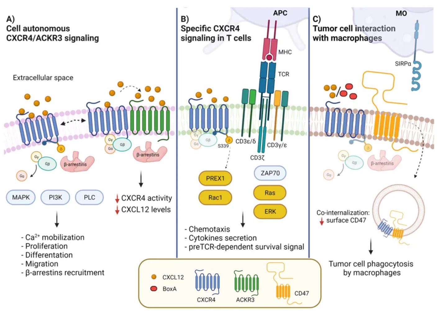
The interactions between the CXCR4 receptor, its ligand CXCL12, and other interacting molecules, along with their roles in cell signaling. Image adapted from: Cancers (Basel). 2022 May 6;14(9):2314.
CXCR4 (C-X-C motif chemokine receptor 4) is a 7-transmembrane helix G protein-coupled receptor encoded by the CXCR4 gene. It participates in various physiological processes and forms crucial interactions with its endogenous ligand, CXCL12 (also known as SDF-1). By binding with CXCL12, the CXCR4 receptor activates multiple signal transduction pathways—including MAPK, PI3K, and PLC—thereby triggering various biological effects such as calcium ion mobilization, cell proliferation, migration, and differentiation. Additionally, CXCR4 can regulate signal transmission by forming heterodimers with ACKR3. In T cells, the interaction between CXCR4 and CD3/T cell receptors (TCRs) promotes the formation of immunological synapses and activates the RAS-ERK signaling pathway. In tumor cells, the interaction between CXCR4 and CD47 promotes their co-internalization, reducing surface CD47 expression and thereby enhancing the phagocytosis of tumor cells by macrophages. CXCR4 plays a key role in the occurrence and development of various refractory diseases, including HIV infection, inflammatory diseases, and metastatic cancers such as breast cancer, gastric cancer, and non-small cell lung cancer. The high expression of CXCR4 in tumor tissues is closely related to tumor invasiveness, metastasis, and increased recurrence risk, making it a focal point for research on CXCR4-targeted imaging and therapy.
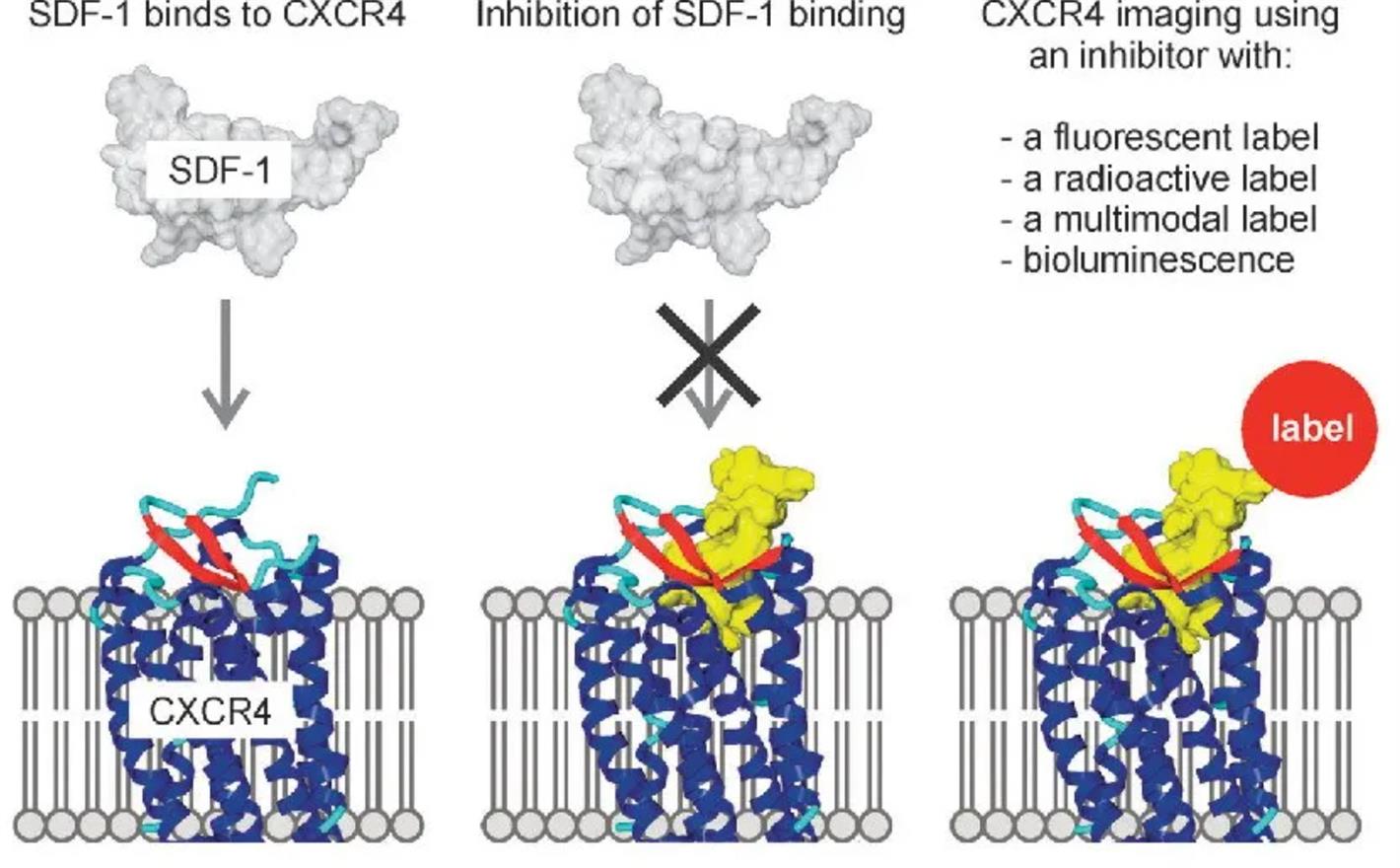 Schematic diagram of the interaction between CXCR4 and SDF-1, and the use of CXCR4 as a therapeutic target (yellow indicates inhibitors) and an imaging target (cropped version). Original image source: Chem Soc Rev. 2012 Aug 7;41(15):5239-61.
The discovery of the CXCR4 inhibitor T140 originated from extended research on HIV infection. Initially, the T22 structure was found to inhibit the infection of T-cell line-tropic HIV-1 by specifically binding to the chemokine receptor CXCR4. This is a peptide amide composed of 18 amino acid residues with potent anti-HIV activity. Subsequently, researchers discovered a more potent and smaller CXCR4 inhibitor 2 (T140; anti-HIV activity: 50%, effective concentration EC50 = 3.5 nM, antagonistic effect on X4-HIV-1 entry: EC50 = 0.43 nM, CC50 = 45 µM). Through in-depth structure-activity relationship (SAR) studies, researchers identified four key amino acid residues (Arg2, Nal3, Tyr5, and Arg14) that are crucial for the activity of the 14-mer peptide. These groundbreaking research results inspired scientists to further optimize the compound structure, ultimately leading to the successful development of FC131, a pentapeptide compound with high affinity for CXCR4. Based on the FC131 structure, CPCR4-2, also known as Pentixafor, was subsequently developed. [68Ga]PentixaFor has become the preferred molecular probe for targeting CXCR4 imaging in the field of nuclear medicine and is increasingly being utilized in Positron Emission Tomography (PET) imaging.
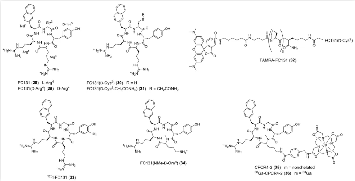 Some relevant chemical structures (partial display shown). Image source: Chem Soc Rev. 2012 Aug 7;41(15):5239-61.
Global R&D Overview of CXCR4 Radiopharmaceuticals
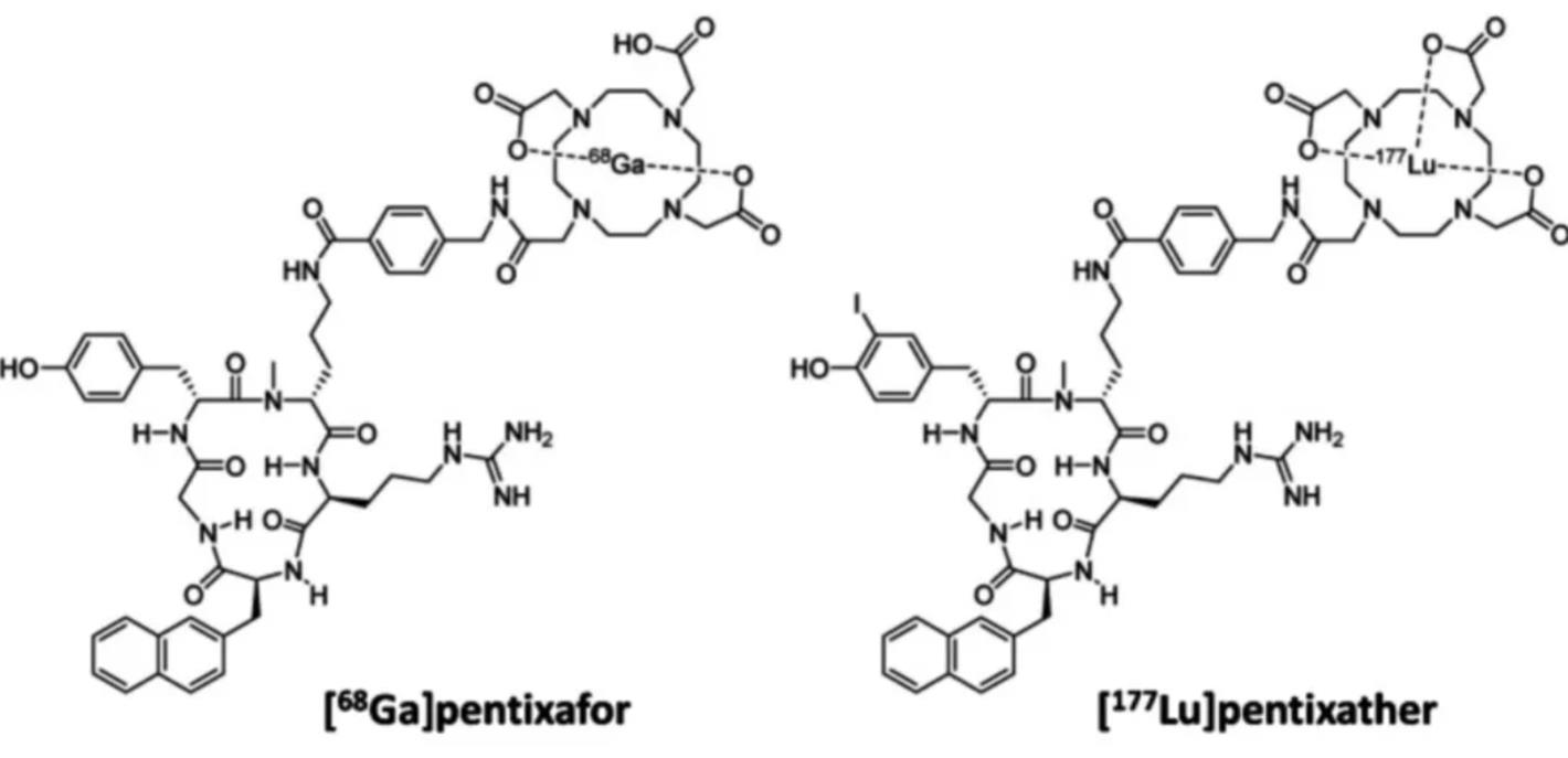 Image source: Eur J Nucl Med Mol Imaging. 2022 Nov;49(13):4616-4641.
Currently, a variety of radiopharmaceuticals are under development worldwide at different stages, primarily focusing on oncology, hematology, rare diseases, and diagnostic agents. Among them, 68Ga-pentixafor, originally developed by PentixaPharm (a wholly-owned subsidiary of Eckert & Ziegler SE) and Scintomics, targets various conditions including marginal zone lymphoma, adrenal diseases, multiple myeloma, and brain cancer. It entered Phase III clinical trials on November 9, 2023.
90Y-pentixather and 177Lu-pentixather, originally developed by PentixaPharm and Technische Universität München as well as Scintomics, are mainly used to treat multiple myeloma, T-cell lymphoma, and indolent non-Hodgkin lymphoma. They entered Phase I/II clinical stages on January 26, 2023, and April 10, 2024, respectively. Additionally, drugs such as 203Pb-pentixather and 212Pb-pentixather, originally developed by the University of Iowa, entered Phase I clinical trials on September 28, 2022. Targeting tumors and rare diseases, they are intended for the diagnosis/treatment of lung carcinoids and small cell lung cancer. [18F]BL40, originally developed by BC Cancer, is applied in oncology, hematology, rare diseases, and diagnostic agents for treating marginal zone lymphoma, multiple myeloma, mantle cell lymphoma, chronic lymphocytic leukemia, and small lymphocytic lymphoma. It entered Phase I clinical trials on January 25, 2024.
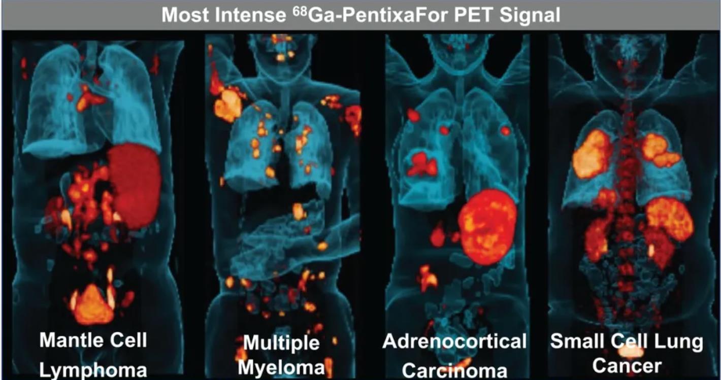 Image source: Journal of Nuclear Medicine November 2022, 63 (11) 1687-1692;
PET imaging with 68Ga-pentixafor has been proven to be highly effective in the diagnosis, subtyping, and staging of diseases such as primary aldosteronism, myeloma, indolent lymphoma, and pituitary tumors. This technology provides a convenient, intuitive, and effective reference for clinical decision-making, aiding in disease prognosis prediction and treatment response assessment.
In a clinical study published in the Journal of Nuclear Medicine in November 2022, 690 tumor patients underwent 777 scans of 68Ga-Pentixafor PET/CT. The results showed that 68.9% of the scans exhibited distinct uptake in disease sites, with particularly high tracer uptake (SUVmax > 12) observed in multiple myeloma, adrenocortical carcinoma, mantle cell lymphoma, adrenocortical adenoma, and small cell lung cancer. Furthermore, tumor-to-blood pool ratio (TBR) analysis revealed significantly elevated TBR values exceeding 8 in multiple myeloma, mantle cell lymphoma, and acute lymphoblastic leukemia. Although no significant correlation between SUVmax and TBR was found in the study, the high-contrast images demonstrated by 68Ga-Pentixafor across various tumors provide a crucial basis for identifying patients who may benefit from CXCR4-targeted therapies.
Recent Studies on Novel CXCR4 Radiopharmaceuticals (Examples)
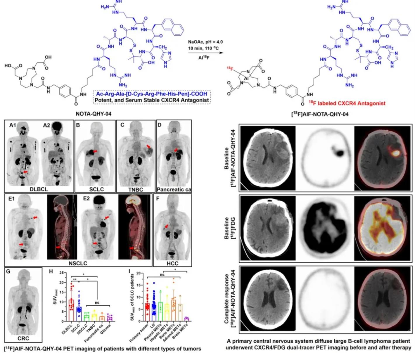 Image source: Journal of Nuclear Medicine June 2024, 65 (supplement 2) 242481;
Conference data from 2024 indicates that researchers have successfully developed a novel CXCR4-targeted PET tracer, [18F]AlF-NOTA-QHY-04, which features high yield, high specificity, and high affinity for CXCR4. Both in vitro and in vivo experiments demonstrated the excellent stability and high binding affinity of [18F]AlF-NOTA-QHY-04 to CXCR4. In PET/CT imaging of murine tumor models and cancer patients, the tracer exhibited tumor uptake that was highly correlated with CXCR4 expression levels.
Furthermore, compared with [18F]FDG, [18F]AlF-NOTA-QHY-04 showed a higher tumor-to-normal tissue ratio in patients with primary brain tumors. Immunohistochemical staining further confirmed that the high tracer uptake was attributed to the high expression of CXCR4 in tumor cells as well as macrophages infiltrating the tumor microenvironment. Therefore, [18F]AlF-NOTA-QHY-04 PET/CT imaging holds potential not only for the precise diagnosis of lymphoma and CXCR4-targeted therapy but also serves as a complementary diagnostic tool to [18F]FDG PET/CT for brain tumors.
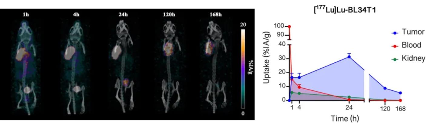 Image source: Journal of Nuclear Medicine June 2024, 65 (supplement 2) 242206;
A 2024 conference report indicates that researchers successfully synthesized a novel CXCR4-targeting peptide, BL34T1. In vitro experiments demonstrated that BL34T1 has high binding affinity for human CXCR4. Furthermore, in vivo studies showed that [177Lu]Lu-BL34T1 exhibited rapid tumor uptake in CXCR4-expressing Patient-Derived Xenograft (PDX) models of Neuroendocrine Prostate Cancer (NEPC), reaching peak tumor uptake after 24 hours.Compared to the previously developed [177Lu]Lu-BL34, [177Lu]Lu-BL34T1 demonstrated longer retention times in the blood, kidneys, and tumors, resulting in a higher tumor absorbed dose. Conjugating the albumin-binding moiety to BL34 did not compromise its CXCR4-specific binding characteristics. The slow clearance of [177Lu]Lu-BL34T1 led to high tumor uptake in CXCR4-expressing NEPC PDX models, thereby extending tumor retention time and achieving a higher tumor absorbed dose, which is beneficial for Radioligand Therapy (RLT).
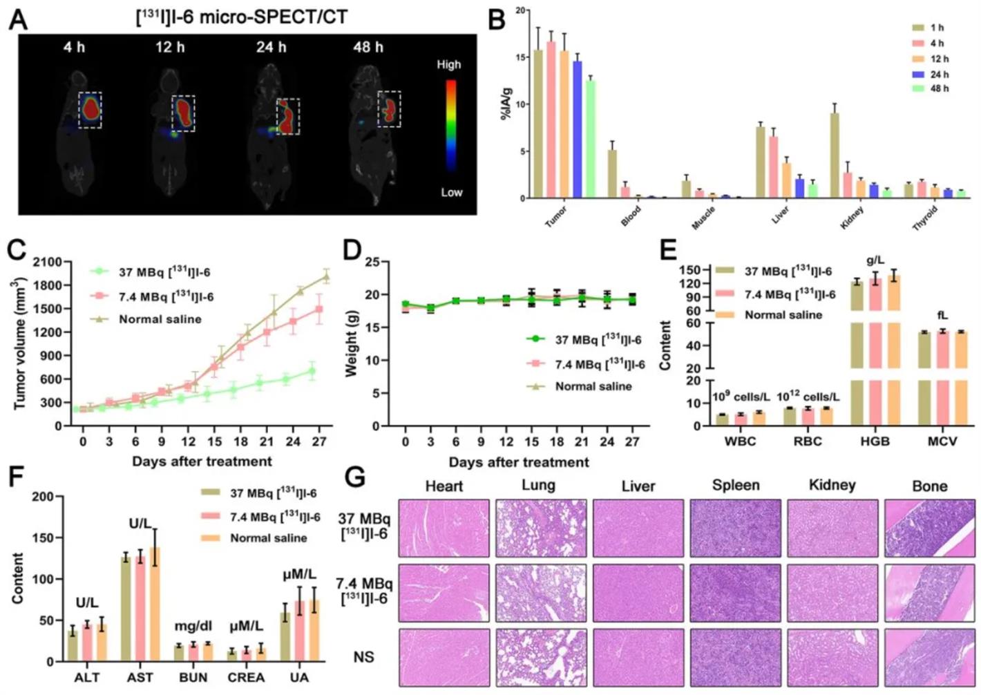
Image source: Journal of Nuclear Medicine June 2024, 65 (supplement 2) 241637;
Another 2024 study focused on the critical role of the chemokine receptor CXCR4 in the development of malignant tumors and explored the potential of integrated nuclear diagnostic and therapeutic approaches targeting CXCR4. The research team screened tumor cell lines with high CXCR4 expression and synthesized six radiolabeled tracers based on the CPCR4 pharmacophore of pentixafor, including [124I]I-1, [99mTc]Tc-2, [124I]I-3, [18F]AlF-4, [99mTc]Tc-5, and [124I]I-6. These tracers were evaluated in animal models via PET/SPECT imaging to identify the most promising candidates for diagnosis and therapy. The results showed that [124I]I-6 possessed the optimal target-to-non-target ratio and was therefore selected as the candidate for therapeutic radionuclide labeling. Following treatment with [131I]I-6 in animal models, the tumor volume in the treatment group was significantly reduced. The study indicates that modifications to molecular probes based on the CPCR4 pharmacophore must be performed cautiously, as changes in chelators or single atoms can significantly impact the probe's affinity and metabolism. [124I]I-6 and [131I]I-6 demonstrated targeting affinity for CXCR4, holding immense potential for the integrated diagnosis and therapy of CXCR4-overexpressing tumors.
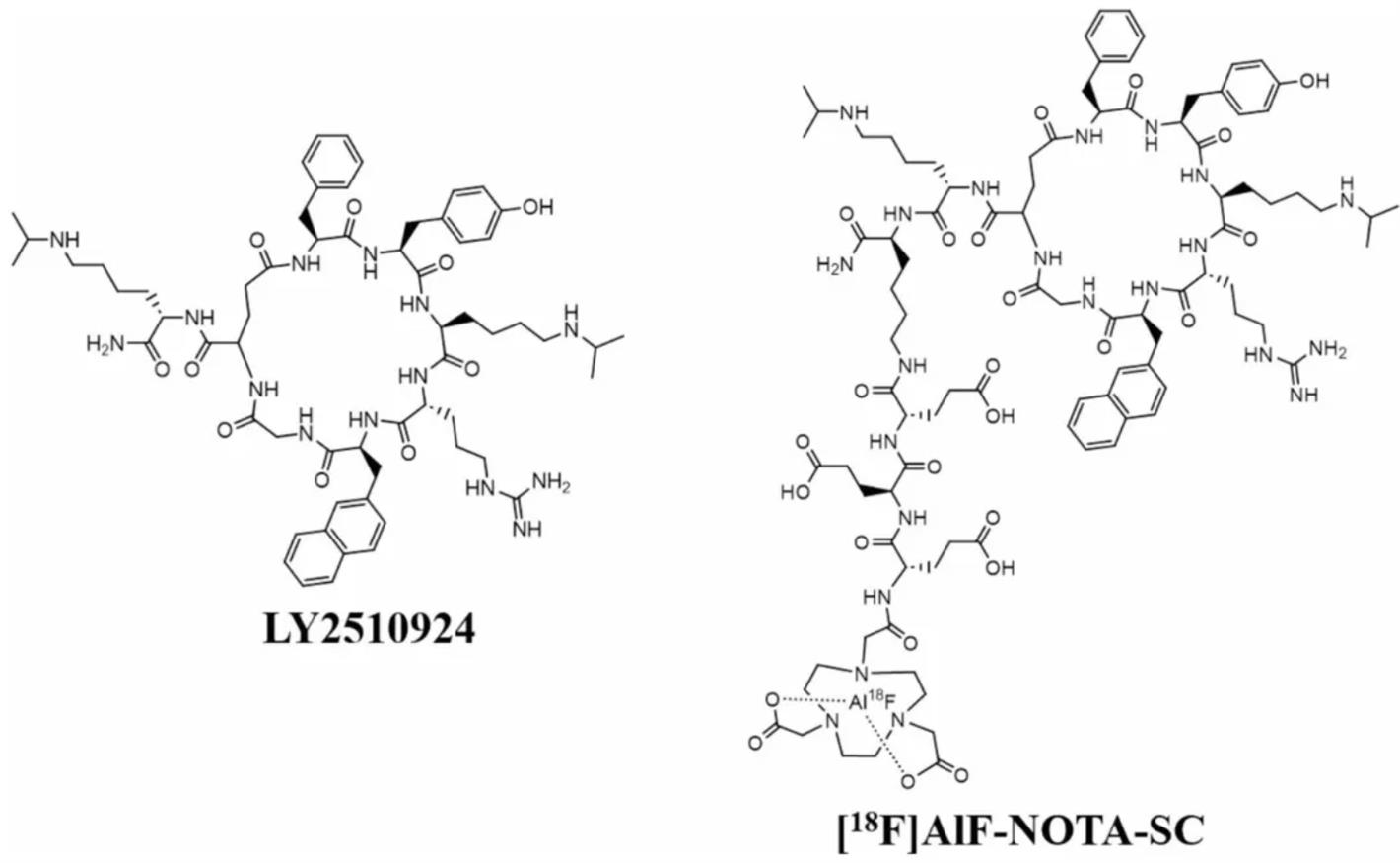 Chemical structures of LY2510924 and [18F]AlF-NOTA-SC. Image source: Eur J Nucl Med Mol Imaging. 2024 Dec 11.
LY2510924 is an orally bioavailable CXCR4 antagonist originally developed by Eli Lilly and Company. It possesses extended in vivo stability and exhibits high affinity for CXCR4 but has been discontinued due to insufficient efficacy. It interacts with the CXCR4 binding site through hydrogen bonding, salt bridges, and π-π stacking.
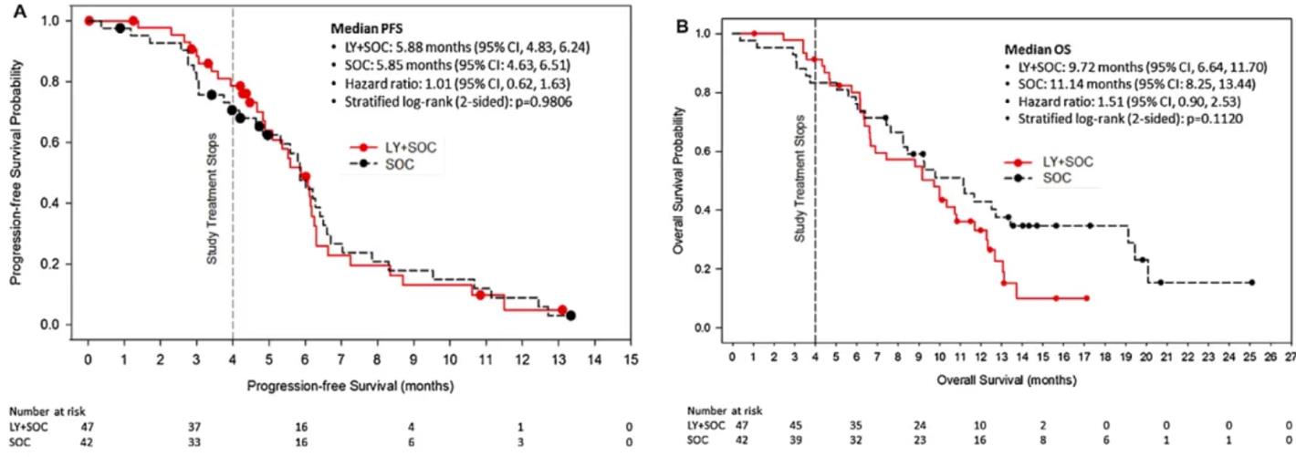 Image source: Lung Cancer. 2017 Mar;105:7-13.
In the treatment of small cell lung cancer, the combination regimen of LY2510924 with carboplatin and etoposide showed suboptimal results in first-line therapy. (ClinicalTrials.gov NCT01439568) This multicenter, open-label, randomized Phase II trial evaluated the efficacy and safety of LY2510924 (LY) as an adjunct to the standard chemotherapy regimen for first-line treatment of Extensive-Stage Small Cell Lung Cancer (ED-SCLC) and explored the potential of C-X-C motif receptor 4 (CXCR4) tumor expression as a predictive biomarker for treatment response. Ninety-four treatment-naïve ED-SCLC patients were randomized 1:1 to receive either carboplatin/etoposide (SOC) alone or in combination with LY2510924 (20 mg, subcutaneous injection, days 1–7 of each cycle) (LY+SOC). The primary efficacy endpoint was Progression-Free Survival (PFS); secondary endpoints included Overall Survival (OS), Overall Response Rate (ORR), and treatment safety. The study results showed no significant difference in median PFS between the LY+SOC group and the SOC group (5.88 months vs. 5.85 months, hazard ratio 1.01, p=0.9806). Median OS also showed no significant improvement (9.72 months vs. 11.14 months). Although the ORR in the LY+SOC group was slightly lower than in the SOC group (74.5% vs. 81%), safety analysis indicated that the addition of LY2510924 did not result in increased toxicity, although the incidence of certain adverse events such as anemia, neutropenia, leukopenia, vomiting, and pneumonia was slightly higher in the LY+SOC group. Furthermore, patients with high baseline CXCR4 expression (H-score 210) did not exhibit a better response to LY2510924 treatment. In conclusion, as an adjunct to first-line therapy for ED-SCLC, LY2510924 failed to enhance efficacy.
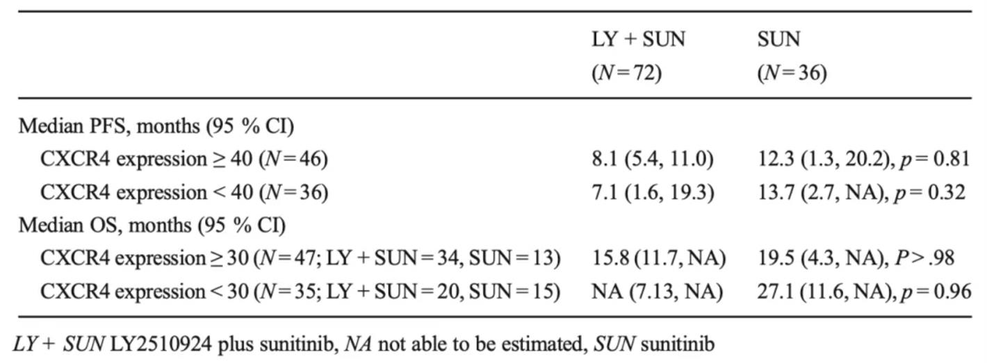 Image source: Target Oncol. 2016 Oct;11(5):643-653.
Regarding renal cell carcinoma treatment, the combination regimen of LY2510924 with sunitinib also showed poor efficacy in first-line therapy. (ClinicalTrials.gov NCT01391130) This randomized Phase II trial evaluated the safety and efficacy of the CXCR4 peptide antagonist LY2510924 (LY) in combination with sunitinib (SUN) as first-line treatment for advanced Renal Cell Carcinoma (RCC). 108 patients were randomized 2:1 to receive either LY (20 mg, subcutaneous injection, once daily) plus SUN (50 mg, oral, once daily, 4 weeks on/2 weeks off) or SUN alone. Treatment continued until disease progression or intolerable toxicity. The study aimed to detect at least a 47% increase in median Progression-Free Survival (PFS). The results showed a median PFS of 8.1 months in the LY+SUN group versus 12.3 months in the SUN alone group. The Bayesian time-to-event hazard ratio was 1.23 (95% CI 0.74–1.96), failing to meet the objectives. LY was well tolerated, and its toxicity profile was consistent with SUN. No significant difference in treatment effect was observed between patient subgroups with high versus low CXCR4-expressing tumors. Therefore, although the addition of LY was acceptable in terms of safety, it did not improve PFS or Overall Survival (OS), indicating that the efficacy of CXCR4 as a therapeutic target in RCC remains unproven.
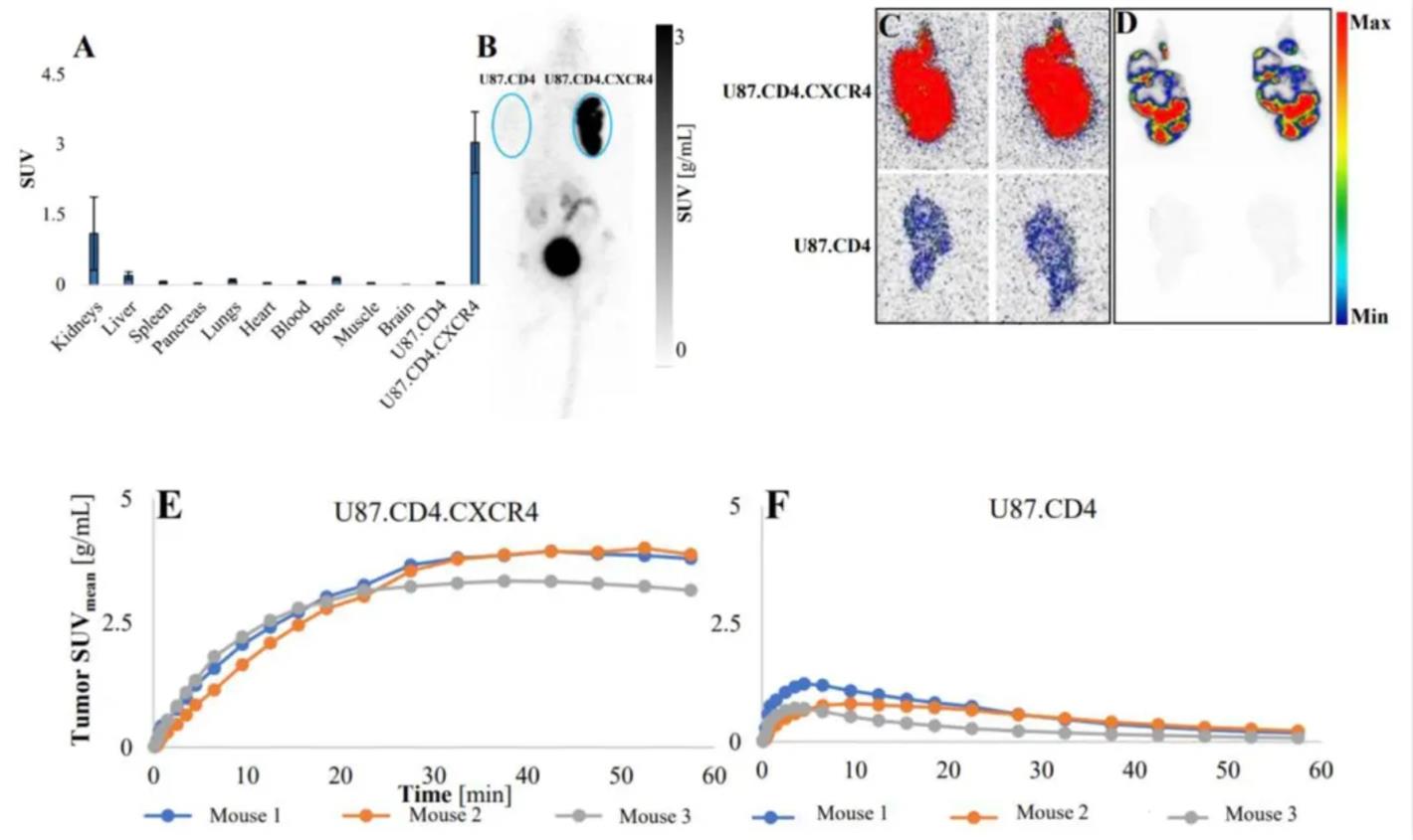
Shows the biodistribution characteristics of [18F]AlF-NOTA-SC in U87.CD4 and U87.CD4.CXCR4 xenograft models, including biodistribution charts, MIP images, and corresponding TAC curves.
Image source: Eur J Nucl Med Mol Imaging. 2024 Dec 11.
A recent study published in the European Journal of Nuclear Medicine and Molecular Imaging aimed to develop an Al18F-labeled radiotracer, [18F]AlF-NOTA-SC, based on LY2510924, utilizing a triglutamic acid linker and NOTA chelator for Al18F labeling. The study results showed that AlF-NOTA-SC exhibits similar in vitro affinity for human CXCR4 compared to Ga-Pentixafor. The decay-corrected radiochemical yield of [18F]AlF-NOTA-SC was 21.0±7.1%, with a specific molar activity of 16.4±3.6 GBq/µmol. In binding assays with U87.CD4.CXCR4 cells, the total binding fraction was 7.1±0.5% (58% blocked by AMD3100). In normal mice, the radiotracer did not accumulate in any organs; however, in tumor-bearing mice, significant CXCR4-specific uptake was observed. In non-human primates, [18F]AlF-NOTA-SC showed uptake in CXCR4-expressing organs such as the spleen and bone marrow. In conclusion, [18F]AlF-NOTA-SC demonstrates CXCR4-specific uptake both in vitro and in vivo, along with rapid and persistent tumor retention, making it a candidate for clinical translation.
Conclusion
The future development of CXCR4-targeted radiopharmaceuticals is exhibiting a trend towards diversification and in-depth research, particularly in the innovation of precursor molecules, expansion of indications, novel applications of radionuclides, and combination therapy strategies. With the deepening understanding of the mechanisms by which CXCR4 functions in tumor development, we anticipate significant breakthroughs in the clinical application of these targeted radiopharmaceuticals, bringing new hope to patients.


Address:Ping An Fortune Center, Lize Financial Business District of Beijing